In April 2008 our practice expanded our Planmeca ProMax to a ProMax 3d, adding Cone Beam CT(CBVT) technology to the unit. The Planmeca ProMax 3D has the ability to work as a regular digital panoramic unit and by switching the sensor it will work as a CBVT although limited to an image size that is 8cmX8cmX8cm, which can be limiting at times.
For the past 2 years we have used the digital pan features of the ProMax for both regular panoramic images and bitewing images. It is a great unit for reading images for perio and does a decent job detecting decay. If it’s a shadow on the Pan image it usually is decay. I like the BiteWing image because it allows me to see the whole tooth during the periodic exam.
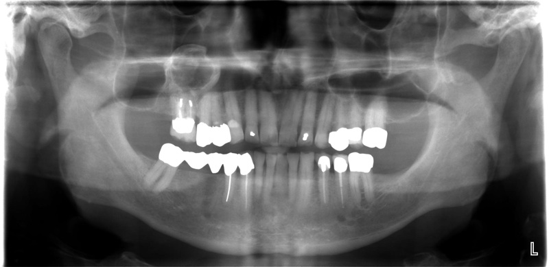
Panoramic Image

Bitewing Image
There are many times I’ll add additional Sensor PA’s if an area isn’t clear. I’ve very comfortable with the diagnostic ability of the Planmeca regular and bitewing panoramic images.
A few questions I had before we upgraded to the CBVT was would I use it for general diagnosis and not just for implant planning?
Would it be a tool that allowed me to find areas of decay that weren’t otherwise noticeable?
Would I be able to see root fractures?
What about early Perio disease?
How would I incorporate it into the exam?
So far I am very pleased with the upgrade to the CBVT and glad we did it, but not as an answer to any of the questions listed above. I love being able to immediately take a scan and be able to see if dental implants are possible? Is grafting necessary? Many times those questions can be answered very easily. I find it’s overkill for decay detection, an explorer and a PA are faster and far less expensive. As far as finding root fractures, I probably wouldn’t extract a tooth just based on the CBVT image, but it’s fun to get a scan to see if you can find the fractured root, and if there were no other symptoms or sequelae it would be hard to find a vertical fracture with a CBVT scan. While it is very easy to see bone loss on the CBVT scan it is over kill also as a perio screening tool in my opinion. A good panoramic or FMX with perio charting is far faster and probably more diagnostic. So far I haven’t seen the need to incorporate it into the comprehensive exam, each image is about 250 megs, so it does take some time for the computers to build the 3d images and it takes time to read the scans.
I think there will be software and resolution improvements down the road that will make the details standout better and hopefully improve it’s limitations for the general practice where dental implants aren’t a major source of treatment.
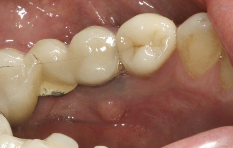
Clinical examination reveals a bump on the lingual mucosa, it has been present for a few weeks and will change in size.
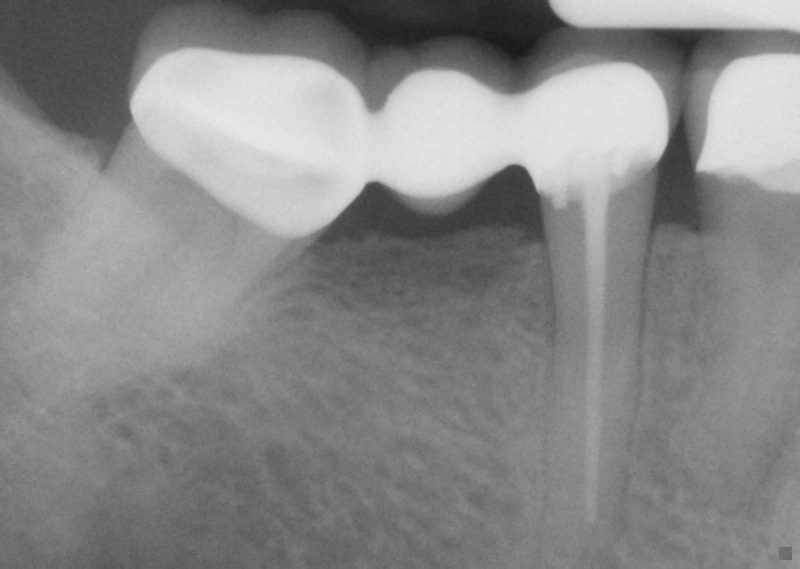
Radiographic evidence shows a halo in the coronal portion of the root.
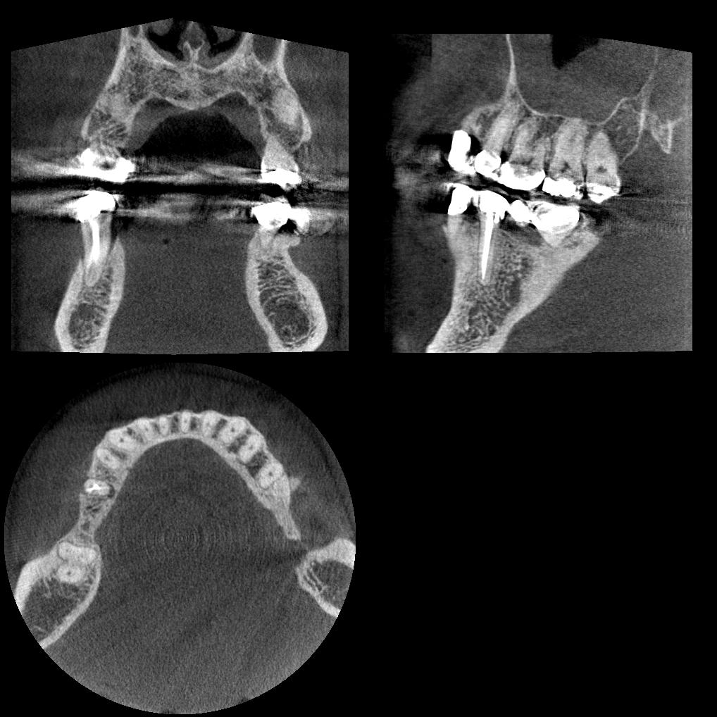
Here’s a 2d image from the Planmeca CBVT. You can see the cross section and see the bone loss on tooth 29. The previous two images are consistent with root fracture, and this image confirms it.. and if I move through the slices with the software I can “almost” visualize the crack, and I’m probably seeing that because I could see the fracture when I extracted the tooth.
Overall I am very pleased with the unit, because we can still take regular panoramic images as well as the occasional CBVT image for implant planning.
Comments on this entry are closed.
Comments
Do you trust the bitewing images to detect and diagnose class 2’s that require treatment vs incipients which do not? Or, are you using more as a screening tool? When you use the bwx option, how do you code for it?
Hi Lee, thanks for your comment. 90+% of my hygiene BWX are used with the Planmeca and I use the BWX code. I see so much more than with digital sensor bwx, you see the whole tooth root and all. When I first got it a few years ago I would take both sensor and planmeca bite wings and rarely found a difference. The reality is that all digital systems do a poor job of detecting inter proximal incipient lesions and I think CR Reports has verified this as true as well. No matter what digital system I use, I check inter proximal areas with my loupes on, a sharp explorer and try to use illumination if possible.